For this task, I have conducted research in to the body’s circulatory system. This research will help me gain a deeper understanding in to how the blood flows around the body, allowing me to create the most realistic wounds possible, in my final Special Effects Makeup assessment.
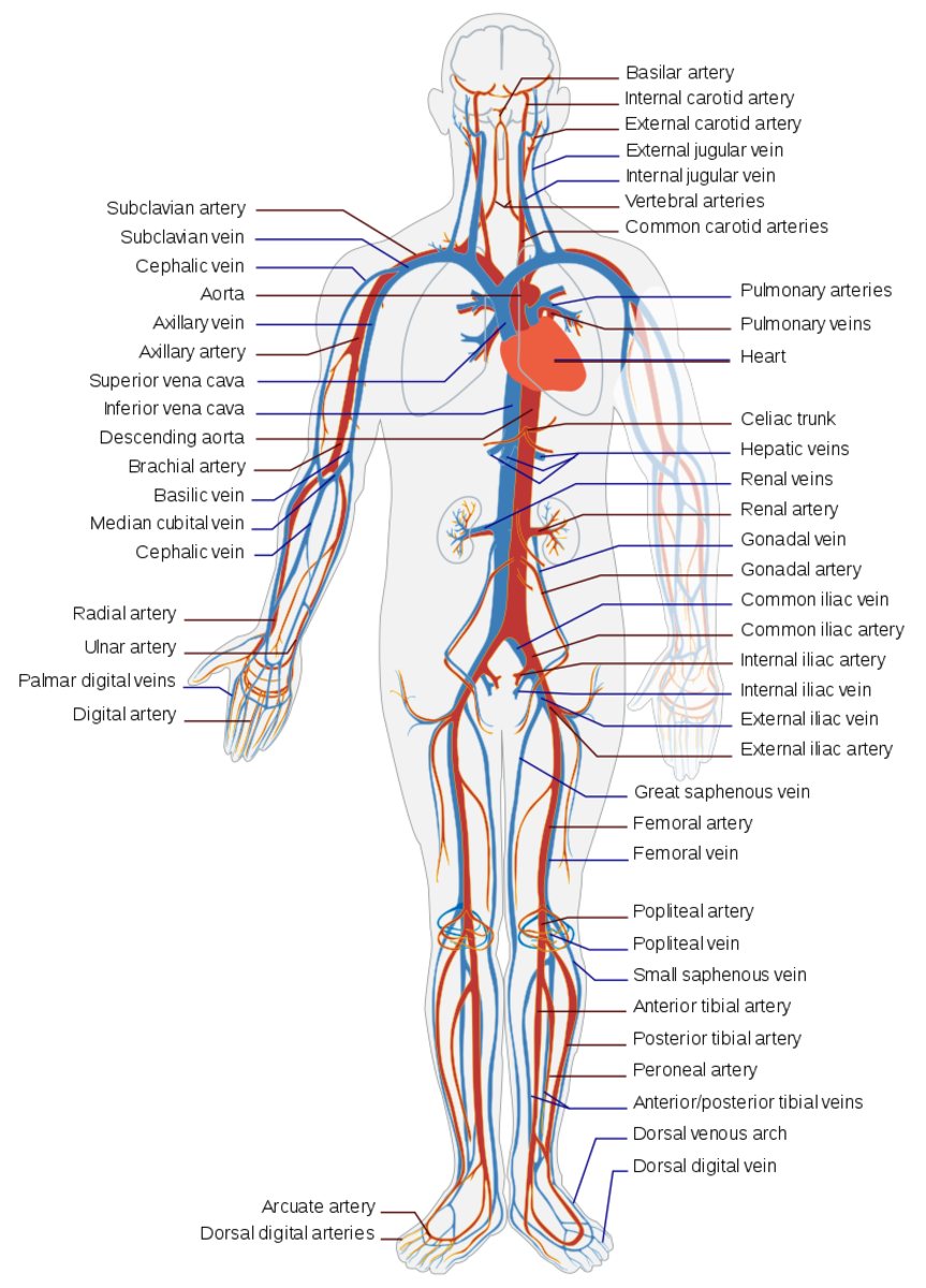
The circulatory system is a system consisting of the heart, blood vessels (arteries, veins and capillaries), and blood. This system is responsible for transporting blood around the body, ensuring tissues receive oxygen and nutrients, and the removal waste products.
How it works;
Step 1 – Oxygen enters the bloodstream, via tiny membranes in the lungs, that absorb oxygen when we inhale.
Step 2 – As the body uses up this oxygen, and processes nutrients in the body, it creates carbon dioxide as a waste product.
Step 3 – This product is then returned to the lungs, and expelled when we exhale.
Constant pressure from the heart and valves, through the body, allows this system to work. This pressure ensures veins can carry blood to the heart, and allows arteries to carry blood away from it. There are three types of circulation, called;
Pulmonary circulation – Where oxygen-depleted blood is carried away from the heart, to the lungs, and then returned back into the heart.
Systemic circulation – Carries oxygenated blood away from the heart, to the rest of the body.
Coronary circulation – Provides the heart with oxygenated blood, allowing it to function properly.
There are many conditions that may potentially affect the effectiveness of the circulatory system. These include;
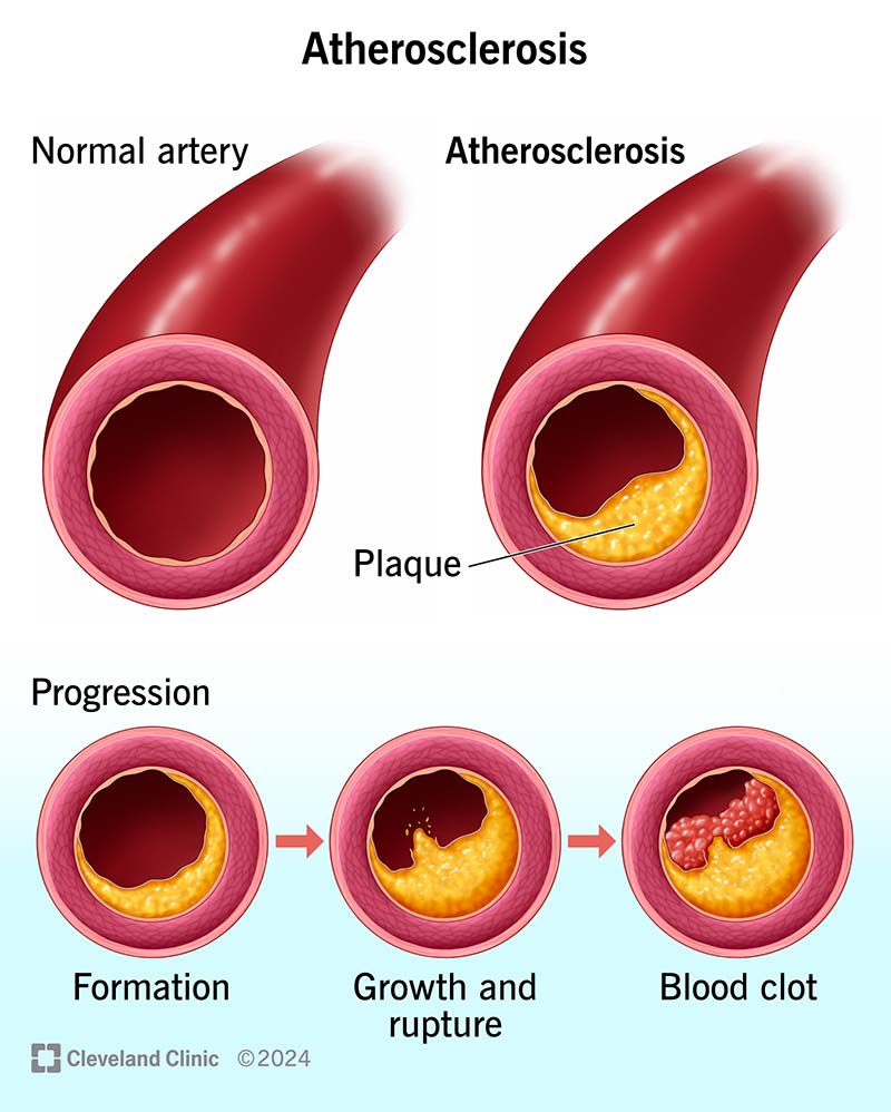
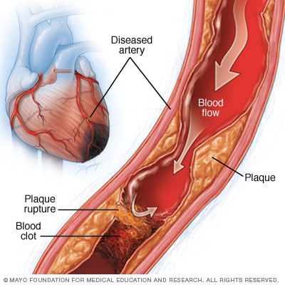
Peripheral arterial disease – A disease where blood flow in the arteries of the leg is restricted, due to a buildup of plaque in the arteries.

Arteriosclerosis – Where a plaque build up in the blood vessel becomes calcified (hardens) causing the arteries to become less flexible. This leads to high blood pressure, and potentially can cause strokes, and heart or kidney damage.
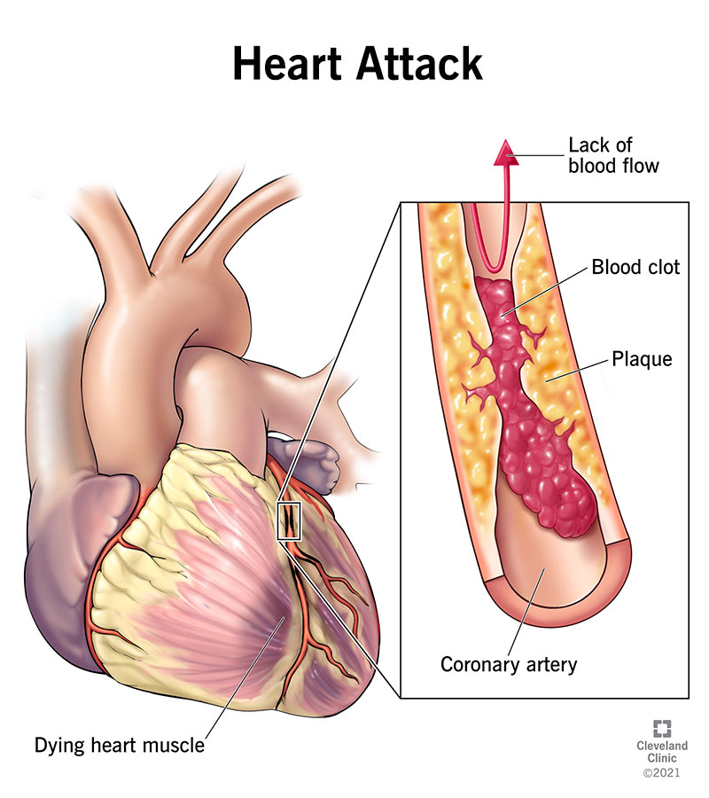
Heart attacks (myocardial infarction) – A blockage of blood flow into the heart muscle, causing the death of muscle tissue in the heart.
Angina – Where the heart muscle doesn’t receive enough blood, causing ‘crushing’ chest pain, fatigue, nausea and shortness of breath.

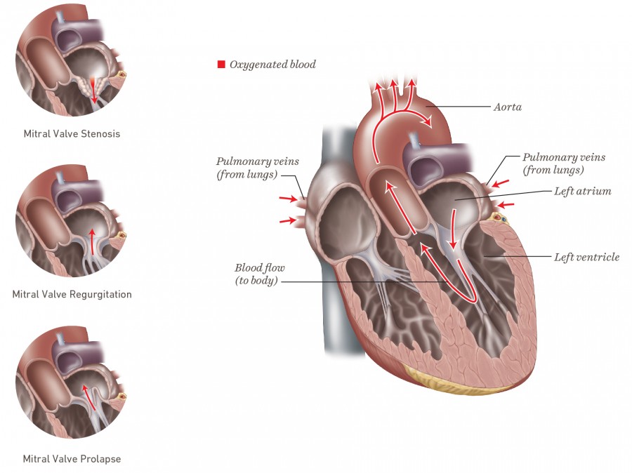
Mitral valve conditions – Known as either mitral valve prolapse, stenosis or regurgitation, these conditions cause oxygenated blood in the heart to flow backwards, slower, or describe is where the blood flow is constricted.
Arrhythmia or dysrhythmia – This term describes an abnormal heart rate.

Ischemia – Where there is not enough blood flow in the heart, meaning the muscles don’t receive enough oxygen.
Aortic disease – A group of conditions affecting the aorta. An example of this is an aneurysm – where the aorta weakens, accusing it to bulge out.

Signs of poor circulation may include ;
Chest pain, Dizziness, Fatigue, Feeling faint, Shortness of breath, Heart palpitations, A slowed or rapid heart beat, Pain, weakness, swelling or numbness in the limbs,
Symptoms specific to conditions, such as Peripheral Arterial Disease include;
Leg cramping – during activity, and rest, Coldness in the feet or legs, A change of color in the leg, Change of nail color or thickness, Hair loss on the legs or feet, Ulcers on the legs or feet that do not heal.
Components
The heart

When fully grown, the heart is around the size of two adult hands. The heart is one of the most important organs in the body – as you can not survive without it – and it sits just off center of the chest. The heart’s consistent pumping is what keeps the circulatory system running which, in turn, keeps us alive.
The heart wall consists of three layers, known as;
The epicardium – Outer layer
The Myocardium – Middle (muscular) layer
The Endocardium – Inner layer
The heart’s chambers are extremely important for the circulatory process. These chambers are known as; The right and left atrium (the upper chambers), and the right and left ventricle (the lower chambers). Right and left atrium are responsible for receiving blood from the veins, while the right and left ventricles push blood back out through the body, away from the heart, through the arteries. This means the ventricles have much stronger myocardial layers, than the atrium.
The Arteries
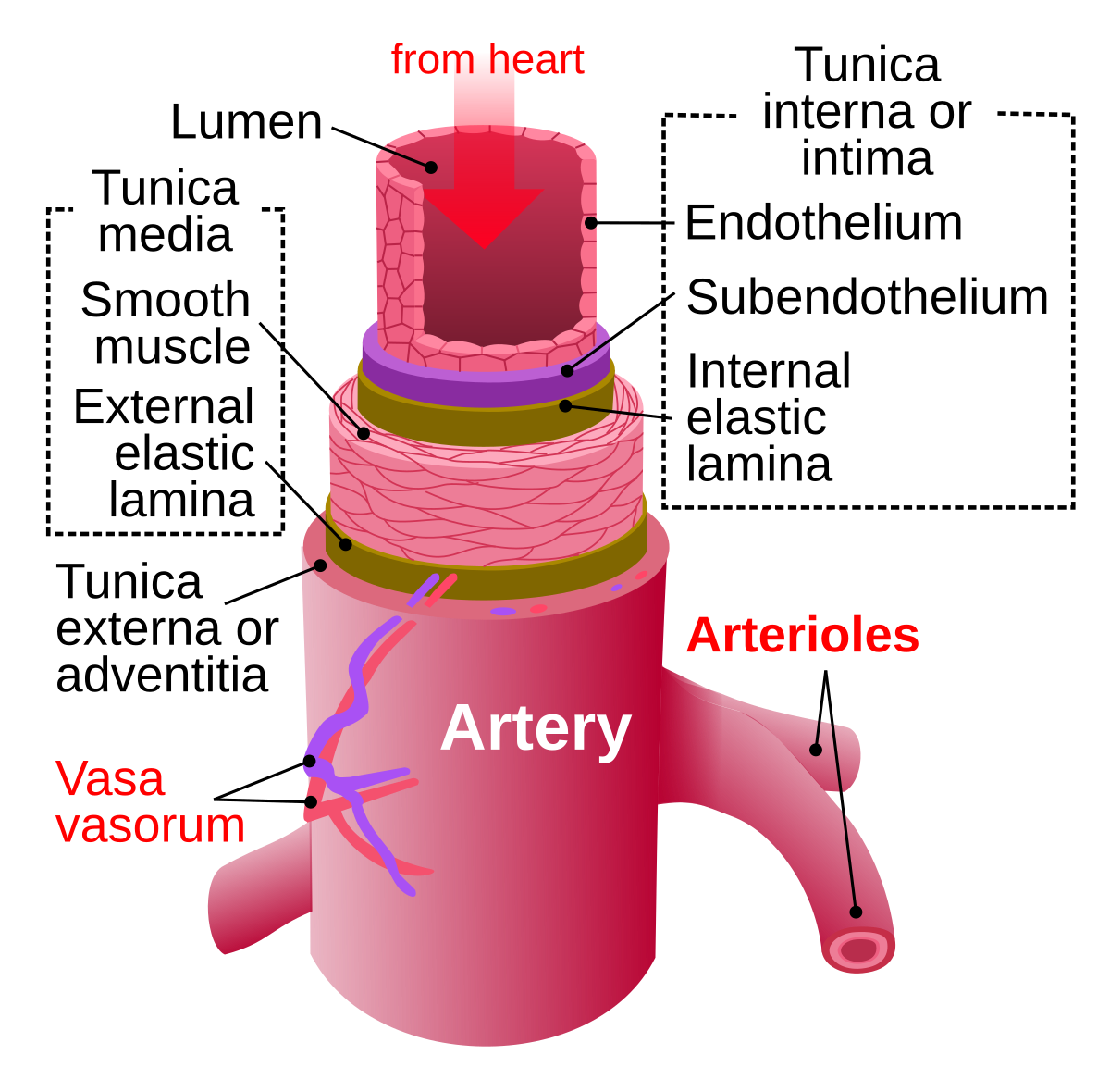
Made of three layers, known as Tunica intima (inner layer), tunica media (middle), and tunica externa (outer layer), the arteries are responsible for transporting blood away from the heart. The tunica media is the thickest of these layers, and changes size to regulate the blood flow.
There are three main types of arteries that get smaller, as they branch further away from the heart.
Elastic arteries;
The aorta and pulmonary arteries are known as elastic arteries. These arteries receive blood straight from the heart, and must be elastic to allow them to accommodate for the surge and contractions, as blood pushes through with each heart beat.
The Aorta, is the largest and most important artery in the body, responsible for carrying blood directly from the heart, into the circulatory system.
The Pulmonary artery is responsible for carrying blood from the right ventricle, to the lungs. This artery is the only artery that can carry de-oxygenated blood.
Muscular arteries;
Muscular arteries are responsible for the movement of blood, between the elastic arteries and the rest of the body. These arteries are made of smooth muscle that expands and contracts as the blood flows. The femoral and coronary arteries are among the most important arteries in this category.
Arterioles;
Arterioles are the smallest of arteries, and are responsible for moving blood from the muscular arteries, to the capillaries.

Capillaries

The capillaries are responsible for connecting the arteries to the veins. The number of capillaries is dependent on the amount of material exchange in an area. For example, skeletal muscle, liver and kidneys contain many capillaries, as their functions require large amounts of oxygen and nutrients, compared to the corneal of the eye, which consists of no capillaries.
Veins
Veins are responsible for the movement of blood, back into the heart. Blood moves through the capillaries to the venules ( the smallest veins ) and then travels its way back to the heart. Just like arteries, the veins get larger in size, the closer they get to the heart, and are also made of three layers, known as ; tunica intima, tunica media, and tunica externa.
However, veins have much less smooth muscle and connective tissue, the walls of veins are much thinner, and they have less pressure, and can hold much more blood, than the arteries.
At any one time, around 70% of our body’s total blood supply is in our veins.
Valves

Valves are small pieces of tissue, found in the veins and the heart, that help to ensure blood is always flowing in the right direction.
Valves can be found in the medium and large veins, in order to help keep the blood flowing toward the heart. Valves, found in veins in the arms and legs, are there to ensure gravity does not pull blood in the wrong direction (away from the heart).
The heart consists of four valves that help to separate the heart’s chambers. There are known as;
Tricuspid valve – Separates the right atrium, from the right ventricle.
Material or Bicuspid valves – Separates the left ventricle, from the left atrium.
The Semilunar valves (Pulmonic or pulmonary, and Aortic valves)
Pulmonic or pulmonary valve – Separates the pulmonary artery from the right ventricle.
Aortic valve – Seperates the aorta from the left ventricle.
The blood
The blood is made up of four main contents, plasma, red blood cells, white blood cells, and platelets.
Plasma;
Around 55% of the blood is made up of plasma. This is the component that makes the blood a liquid.
Plasma is responsible for transporting blood cells around the body (through the circulatory system) as well as carrying hormones, nutrients, antibodies and waste products around the body.
The blood’s plasma is made up of; water, salts, sugar, fat and protein.
Red blood cells (erythrocytes) ;

Around 40-45% of the blood’s volume is made up of red blood cells. These cells have no nucleus, making it easy for them to change shape, allowing them to move swiftly through the veins and arteries.
Red blood cells contain hemoglobin (a protein) that carries oxygen from the lungs, to the rest of the body, and returns carbon dioxide to the lungs – where it is then exhaled.
White blood cells (leukocytes) ;

White blood cells make up only 1% of the blood volume, and are responsible for protecting the body from infection. There are five types of white blood cells, known as;
Monocytes – These have a longer lifespan than most white blood cells, and are used to break down bacteria.
Lymphocytes – These cells create antibodies, allowing the body to fight against bacteria, viruses, and any other potentially harmful bodily invaders. There are two types known as B cells ( cells that produce antibodies) and T cells ( Cells that regulate immune cells, and target infected cells and tumors).
Neutrophils – These are the most numerous types of white blood cells, and are responsible for killing and digesting bacteria and fungi. Neutrophils are the body’s first line of defense when an infection strikes.
Basophils – These are the smallest of white blood cells, and are responsible for sounding an alarm when potentially infectious agents invade the blood. These cells also secrete chemicals in response to alarm, and help control the immune system.
Eosinophils – These cells attack and kill parasites, cancer cells, and aid in allergic responses.
Platelets (thrombocytes) ;

Platelets are cell fragments that are essential for the blood clotting process. Platelets stick to injured blood vessel linings, to provide the basis for a clot – which stops bleeding and promotes healing.
